English
Arabic/арабский
Bengali language/বাংলা ভাষার
German/Deutsch
English/English
Chinese/中文
Spanish/Español
Persian/فارسی
French/Français
Hindi/हिन्दी
Indonesian/Bahasa Indonesia
Italian/lingua italiana
Japanese/日本語
Korean/한국어
Polish/Polski
Portuguese/Portugués
Swedish/svenska
Thai/ภาษาไทย
Turkish/Türk dili
Ukrainian/Українська Мова
Urdu/اردو
Vietnamese/Tiếng Việt
Russian/русский язык
[log in Register]

Chordate

dnaoodb: professional biology database , biology encyclopedia

In biology, a Chordate (/ˈkɔːrdeɪt/ Latin:Chordata /kɔːr ˈdeɪtə/ ) It is the highest phylum in the animal kingdom, and it is also the most successfully developed one. Their common feature is that they have notochord, dorsal neural tube and branchial slits (the three major characteristics of chordates) in the whole process of individual development or at a certain period.

Chordates include urochordates, cephalochordates, and vertebrates. In addition to the above main features, chordates also have some secondary features: a closed circulatory system (except Uurochodates), the heart, if present, is always located on the ventral surface of the digestive canal; A certain stage of life history or present throughout life; endoskeleton with germ layer formation. As for the posterior mouth, bilateral symmetry, three germ layers, true coelom and segmental characteristics are also possessed by some invertebrates.

Chordate fossils have been found from as early as the Cambrian explosion, 539 million years ago. Cladistically (phylogenetically), vertebrates – chordates with the notochord replaced by a vertebral column during development – are a subgroup of the clade Craniata, which consists of chordates with a skull. Of the more than 81,000 living species of chordates, about half are ray-finned fishes that are members of the class Actinopterygii and the vast majority of the rest are tetrapods (mostly birds and mammals).

Scientific classification

Alias:
Chordate,Chordata
Latin:
Chordata
Domain:
Eukaryota
Kingdom:
Animalia

Definition

A chordate is an animal that belongs to the phylum Chordata, which is part of the Deuterostomes kingdom. Organisms in the Deuterostomes kingdom have a distinct characteristic: their anus develops before their mouth in early embryonic stages. The phylum Chordata includes a wide range of organisms, as it is comprised of all vertebrates, which are organisms with a backbone, and many invertebrates; organisms that don’t have a backbone. There are three subphyla to Chordata: Cepahlochordata, Urochordata, and Vertebrata. Chordates generally have bilaterally symmetric bodies, though a few exceptions exist, and they share distinct characteristics that will be discussed later on.

History

History of name

Although the name Chordata is attributed to William Bateson (1885), it was already in prevalent use by 1880. Ernst Haeckel described a taxon comprising tunicates, cephalochordates, and vertebrates in 1866. Though he used the German vernacular form, it is allowed under the ICZN code because of its subsequent latinization.

history of zoology

Chordates undoubtedly descended from invertebrates. Among them, echinoderms and hemicordates are closely related to chordates, which is accepted by most people.

It is speculated that the ancestor of chordates may be similar to the larvae of Uurochodates. It developed in two directions, one is through metamorphosis, adults live sessilely, with gill slits as feeding and breathing organs; the other direction is the extension of larval stage And adapt to the new living environment, stop metamorphosis, produce gonads and reproduce (that is, larval sexual maturity), and then develop a new class of animals, that is, free-moving chordates with notochords, dorsal neural tubes and gill slits; after that Differentiate into jawed (fish ancestors) and jawless.

In 2023, an international scientific research team discovered the "Burgess Shale-type" specific buried fossil bank in the Middle Ordovician strata of Wales, England-the Castle Beach Biota, and found more than 170 kinds of organisms, including chordates.

Characteristics

All chordates possess, at some point during their larval or adult stages, five synapomorphies, or primary physical characteristics, that distinguish them from all the other taxa. These five synapomorphies include a notochord, dorsal hollow nerve cord, endostyle or thyroid, pharyngeal slits, and a post-anal tail. The name “chordate” comes from the first of these synapomorphies, the notochord, which plays a significant role in chordate structure and movement. Chordates are also bilaterally symmetric, have a coelom, possess a circulatory system, and exhibit metameric segmentation.

In addition to the morphological characteristics used to define chordates, analysis of genome sequences has identified two conserved signature indels (CSIs) in their proteins: cyclophilin-like protein and mitochondrial inner membrane protease ATP23, which are exclusively shared by all vertebrates, tunicates and cephalochordates. These CSIs provide molecular means to reliably distinguish chordates from all other Metazoa.

notochord

a rod-like structure between the alimentary canal and the neural tube, with a supporting function, all chordates have the notochord in the embryo but are retained later in life or throughout life, or Degenerates and is replaced by the spine .Notochord and Spinal Cord in a Vertebrate
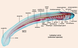
The notochord originates from the dorsal wall of the gastrula during the embryonic period, that is, the notochord mesoderm. After thickening, differentiation, protruding, and finally breaking away from the gastrogut to form the notochord. The notochord is composed of vacuolar-rich notochord cells surrounded by a connective tissue notochordal sheath secreted by notochord cells. The notochord sheath usually consists of two inner and outer layers, a fibrous sheath and an elastic sheath. The notochord cells filled with vacuoles generate turgor and pressure, making the whole notochord both elastic and rigid, thus playing the basic role of bones. In lower chordates, the notochord exists throughout life or is only seen in the larval stage. In higher chordates the notochord appears only during the embryonic period and is replaced by a segmented bony vertebral column when fully developed. The cells that make up the endoskeleton, such as the notochord or spine, grow continuously as the animal body develops. Invertebrates lack endoskeleton such as notochord or spine, usually only the body surface is covered with exoskeleton such as chitin. The appearance of the notochord is of great significance in the history of animal evolution. appears in:

①The notochord (and the spine) constitute the main beam supporting the body, which bears the weight of the body and provides strong support and protection for the internal organs.

②Exercise muscles obtain a strong fulcrum, so that the body will not be shortened or deformed due to muscle contraction during exercise, thus developing towards "larger size". At the same time, the support function of the central axis of the notochord can also enable the animal body to complete directional movement more effectively, and it is more accurate and rapid for active predation and evasion of predators.

③ The formation of the vertebrate skull, the appearance of jaws, and the protection of the central nervous system by the spinal canal are all further perfect developments on this basis.

dorsal tubular nerve cord

The hollow, tubular central nervous system located on the back of the notochord. In vertebrates, the front of the neural tube expands to form the brain, and the back of the brain forms the spinal cord.

Formed by invagination of the ectoderm in the dorsal mid-section of the embryo body. The dorsal neural tube differentiates anteriorly and posteriorly into the brain and spinal cord in higher species. The neurocoele forms the cerebral ventricle in the brain and the centralcanal in the spinal cord. The central part of the nervous system of invertebrates is a solid ventral nerve cord located on the ventral surface of the digestive tract.

The dorsal hollow nerve cord

The dorsal hollow nerve cord is a hollow tube derived from the ectoderm during the embryonic stage of vertebrates. It lies dorsal to the notochord. Thus, it may be seen at the top of the notochord in chordates. This tube is made up of the nerve fibers that ultimately develop into the central nervous system where the brain and the spinal cord are the main constituents. The dorsal hollow nerve cord is protected by the vertebral column.

The nerve cord, though, is not an exclusive feature of chordates. It is also present in other animal phyla. In other animals, it is located either ventral or laterally as opposed to that of chordates that lie dorsal to the notochord.

gill slit

A series of fissures arranged in pairs on both sides of the pharynx, directly or indirectly communicated with the outside world. The gill slits of lower chordates and fish exist throughout their lives, while other vertebrates have gill slits only in the embryonic stage. Derived from ectoderm and endoderm

There are a series of slits arranged in pairs on both sides of the pharynx at the front end of the digestive tract in chordates such as basalis, which directly open on the body surface or indirectly communicate with the outside world through a common opening. These slits are the pharyngeal slits. The gill slits of lower aquatic chordates are present throughout life and are accompanied by vascularized gills. As a respiratory organ, terrestrial higher chordates only have branchial openings in embryonic or larval stages (such as amphibian tadpoles), and eventually die out completely with growth and development. The gills of invertebrates are not in the pharynx, and the organs used for respiration include the pectin gills of molluscs and the limb gills, tail gills, and trachea of arthropods.

Post-Anal Tail

This is a posterior elongation of the body that helps propel aquatic animals in water, provides balance, and is used by some terrestrial vertebrates to attract mates and signal when danger is near. This tail shrinks in humans and other apes into a tailbone during embryonic development.

Circulatory system

Located on the ventral surface of the digestive tract, the circulatory system is closed tube. The heart and aorta of invertebrates are at the back of the digestive tract, and the circulatory system is mostly open tube.

Among the above-mentioned features, the notochord, dorsal neural tube, and pharyngeal branchial slit are the three most important basic features that distinguish chordates from invertebrates. In addition, chordates also have some traits that are also seen in higher invertebrates, such as three germ layers, posterior mouth, existence of secondary body cavity, bilaterally symmetrical system, segmentation of body and certain organs, etc. These commonalities suggest that chordates evolved from invertebrates.

Urochordata

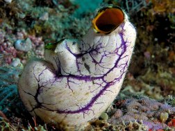
Body wall: The body wall of the sea squirt is the mantle that houses the internal organs. In addition to the epithelial cells of the epidermal layer on the surface of the mantle, it is also mixed with muscle fibers derived from the mesoderm to control the body and enter and exit the water. Expansion and opening and closing of holes. The body wall can secrete a tunicin whose chemical composition is similar to plant cellulose, and thus form a tunic that surrounds the animal body, which is the origin of the name tunicate. In the entire animal kingdom, the animals whose body wall can secrete tunicin have only been found in uronchorates and a few protozoa. The mantle meets the tunicate at the edge of the water inlet and outlet holes, and there is an annular sphincter at the junction to control the opening and closing of the tube holes. Of the internal organs, only the upper edge of the pharynx and a part of the ventral surface were healed with the mantle. The surface of the tunic of sea squirts is usually not easily attached by other animals, but individuals of the same species can overlap, which is obviously positive for the reproduction of the population.Urochordata

Digestive and respiratory system: There is a mouth at the bottom of the water inlet hole, and the wide pharynx passes through the limbal membrane with tentacles around it. The pharynx occupies almost half of the body (3/4), and the pharyngeal wall is covered by many small gill slits. run through. The water that enters the pharynx from the mouth passes through the gill slits, reaches the peribranchial cavity surrounding the outside of the pharynx, and is discharged through the orifice. The peribranchial cavity is a cavity formed by the body surface sinking into the interior. Because of its continuous expansion, the original body cavity in the front of the body is gradually squeezed, and finally disappears completely in the pharynx. Since there are abundant capillaries distributed in the gap between the gill slits, when the water flow carries food particles through the gill slits, gas exchange can be carried out to complete respiration. There are cilia on the inner wall of the pharyngeal cavity, and there is a groove-like structure in the center of the dorsal and ventral sides, which are called dorsal lamina or epipharyngeal groove and endostyle respectively, and there are gland cells and ciliated cells in the groove; the dorsal lamina and The inner columns face each other up and down, and are connected by the peripharyngeal groove at the front end of the pharynx. Gland cells can secrete mucus to make the food that sinks into the inner column stick together into balls, and the cilia in the grooves swing to push the food balls from the inner column It moves forward, passes through the peripharyngeal groove and guides backward along the back plate into the esophagus, stomach and intestines for digestion. The intestines open in the peribranchial cavity, through which indigestible residues are excreted with the water through the outlet holes.

Circulation mode and excretory organs: The heart is located in the pericardial cavity on the ventral surface of the body near the stomach, and beats by the expansion and contraction of the pericardium. There is a blood vessel at each end of the heart, and the front one is a gill blood vessel, which branches along the abdomen of the pharynx to the pharyngeal wall between the gill slits; the rear end is called an intestinal blood vessel, which branches to each internal organ and injects blood into the blood sinus of the organ tissue Therefore, it is an open-tube blood circulation. Sea squirt has a special reversible blood circulation flow direction, that is, the heart contraction has periodic intervals. When its front end continuously beats, the blood is continuously pressed out from the gill blood vessels to the gills, and then the heart has a short pause to accommodate the gills. The blood flows back to the heart, and then begins to beat at the back end, injecting blood into the intestinal vessels and distributing it among the tissues of the internal organs. Therefore, the blood vessels of sea squirts have no distinction between arteries and veins, and the blood has no fixed one-way flow direction. This unique blood circulation mode is unique in the animal kingdom.

Sea squirts have no specialized excretory organs, only a bunch of cells with excretory function near the intestines, called small renal vesicles, which often contain uric acid crystals.

Nerves: Adults of Ascidian stipe live sedentarily, with degenerated nervous system and sensory organs. The central nervous system is just a ganglion (nervus ganglion) without inner cavity, round and hard, shaped like nodules, located at the water inlet and outlet In the mantle wall between the foramen, several nerves branch out to various parts of the body. There is a colorless, transparent and slightly enlarged neural gland (neural gland, which is equivalent to the hypophysis of higher animals) next to the ganglion. No special sensory organs, only a few scattered sensory cells on the limbal membrane and mantle of the inlet and outlet pores.

Reproductive system: hermaphroditic, the gonads are located between the intestinal rings and on the inner wall of the mantle. The testis is large and branched, and it is a milky white granule; the ovary is long tubular, pale yellow, and contains many round egg cells; the two are closely overlapped, and the mature sex cells are separated by a single gonoduct It enters the gill cavity, and then discharges out of the body through the outlet pipe hole, or meets and fertilizes with the germ cells of another sea squirt in the gill cavity.

Cephalochordata

Cephalochordate: Lancelet

Skin: The skin is thin and translucent, consisting of an epidermis of single-layer columnar cells and a dermis of jelly-like connective tissue, and the epidermis is covered with a layer of cuticle. The epidermis has cilia in the larval stage and disappears after growth.

2. Skeleton: Amphioxus has not yet formed a bony skeleton, and mainly uses the notochord running through the whole body as the central axis support for the animal body. There is a notochord sheath around the notochord, which is continuous with the adventitia of the dorsal neural tube, the septum between the sarcomeres, and the subcutaneous connective tissue. The notochord cells are flat disk-shaped, and their ultrastructure is similar to the muscle cells of bivalve molluscs, which can increase the stiffness of the notochord during contraction. In addition, the tentacles of the mouth lip, limbal tentacles, and discs are also supported by horny substances, and the fin rays and gill bars of the odd fins and gill slits are supported by connective tissue.

Muscles: The back muscles of amphioxus are thick and the abdomen is relatively thin, which is different from that of invertebrates where the body wall is uniform in thickness. The main muscles of the whole body are more than 60 pairs of relatively primitive "V"-shaped myomere arranged in sections on the side of the body, with the tip facing forward, and the myomere is separated by the myocomma of connective tissue. The sarcomeres on both sides are asymmetrical, which facilitates the bending movement of amphioxus in the horizontal direction. In addition, there are transverse muscles distributed on the ventral surface of the peribranchial cavity and sphincters on the oromal membrane, etc., which control the drainage of the peribranchial cavity and the size of the orifice.

Digestive and respiratory organs: amphioxus depends on the swing of the wheels and pharyngeal cilia, so that the water with food particles flows into the pharynx through the mouth, the food is filtered and left in the pharynx, and the water passes through the gill slits of the pharyngeal wall to the pharynx. Surrounds the gill cavity, and then exits the body through the ventral opening. The pharynx, which serves as a place for food collection and breathing, is extremely enlarged, occupying almost 1/2 of the total length of the body. The structure of the pharyngeal cavity is similar to that of Uurochodates, and it also has internal columns, superior pharyngeal grooves, and peripharyngeal grooves. The gill slits of amphioxus larvae open directly on the body surface, and later form a peribranchial cavity, with the ventral opening as the main water outlet of the gill slit in the pharynx. This feeding method is passive filter feeding.

The food particles in the pharynx are bound into groups by the secretion of the inner column cells, and then the ciliary movement makes them flow from the back to the front, pass through the peripharyngeal groove to the superior pharyngeal groove, and push them back into the intestine. The intestine is a straight canal, protruding forward into a caeca, protruding into the right side of the pharynx, called the hepatic diverticulum, which can secrete digestive juice, and is the same organ as the liver of vertebrates. The small particles in the food bolus can enter the hepatic caeca, be swallowed by the cells of the hepatic caeca, and be digested in the cells. After the large particles are decomposed into small particles in the intestine, they are also transferred to the hepatic caeca for intracellular digestion. Substances in the hepatic caecum return to the intestine, where they are digested and absorbed in the hindgut. The intestine ends at the anus on the left side of the body.

The pharynx is the part where amphioxus completes respiration. There are more than 60 pairs of gill slits on both sides of the pharyngeal wall, which are separated by gill bars. The inner wall of the gill slits is covered with ciliated epithelial cells and blood vessels. When the water enters the mouth and pharynx, it passes through the gill slit through the movement of the cilia of the ciliated epithelial cells, and makes it exchange gas with the blood in the blood vessel. Finally, the water is discharged from the peribranchial cavity through the ventral hole. Some people think that the lancelet's thin skin also has the ability to absorb oxygen directly from the water.

Blood circulation The circulatory system of amphioxus belongs to the closed-tube type, that is, the blood flows completely in the blood vessels, which is basically the same as that of vertebrates. There is no heart, but the ventral aorta has the ability to beat, so it is called a narrow heart animal. Many pairs of branchial arteries are branched from the abdominal aorta to both sides and enter the branchial septum. The branchial arteries are no longer divided into capillaries. After completing the respiration of gas exchange, they merge into two dorsal arteries at the back of the branchial slit. aortic root. The root of the dorsal aorta contains oxygenated blood, which flows forward to the front of the body, and backward, the dorsal aorta is formed by the roots of the left and right dorsal aorta, and then blood vessels branch out to all parts of the body. The blood is colorless, and there are no blood cells and respiratory pigments. The blood in the arteries enters the veins through the interstitial space. The blood returning from the front of the body is injected into a pair of anterior cardinal veins through the parietal vein; there is a caudal vein on the ventral surface of the tail, which collects part of the blood returning from the back of the body and enters the lower intestine vein (subintestinal vein), most of the blood flows into the 2 posterior cardinal veins (posterior cardinal vein). The blood from the left and right anterior cardinal veins and the two posterior cardinal veins all flow into a pair of transverse common cardinal veins, or ducts Cuvieri. The junction of the left and right common main veins is the sinus venosus, which then leads to the abdominal aorta. The blood returned from the intestinal wall is collected into the inferior intestinal vein by the capillary network, and part of the blood of the tail vein is also injected into it; Capillaries are formed in the caeca area, so it is called the hepatic portal vein (hepatic portal vein). The hepatic vein is formed again from the capillaries of the hepatic portal vein and drains into the sinus venosus.

Subphyla Of Chordates

This is the subphylum that we, and all other vertebrates, belong in. Vertebrata is often referred to as Craniata because the organisms in this subphylum have a head with a protective cranium, which most of us call a skull. Vertebrates also have a brain encased in the skull, highly developed internal organs, a closed circulatory system, and unique sensory and motor cranial nerves. Fish, amphibians, reptiles, mammals, and birds are all part of his subphylum, and thanks to the presence of bone or cartilage, their fossils are easily found.

Subphylum Cephalochordata

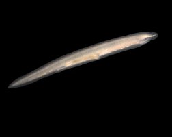
This literally means “head cords”. Cephalochordates are also called Lancelets, and they are filter-feeding marine animals with elongated, small, segmented, and soft bodies that make it difficult to find their fossils. They are usually found in soft bottoms as they bury themselves in the substrate, exposing their anterior part only (near the head) and using a row of tentacles to bring food into their mouth. Cephalochordates resemble fish, as we can see in the picture below, and swim like them, but they have no scales, backbone, limbs, or brain. They outdo fish, however, in the number of pharyngeal gill slits that they have.

Subphylum Urochordata

Meaning “tail cords”, these are also called Tunicates. This is another subphylum whose organisms’ fossils are difficult to find, since the bodies have no hard parts. It includes sea squirts and sea salps. These are barrel-shaped, non-segmented filter-feeding marine animals. The larval stage usually has a tail and is free-swimming, but it eventually attaches to a hard substrate and loses the tail as it transforms into the adult form. By the time these organisms are adults, they only display one characteristic of chordates, and that is pharyngeal gill slits.

Reproduction And Life Cycle

The whole new and diverse class of vertebrates with a vertebral column, tunicates, and lancelets are represented by the phylum Chordata. Two strategies are used for sexual reproduction: 

(1) internal fertilization and 

(2) external fertilization. In the process of internal fertilization, the sperm and the eggs (collectively known as the gametes) unite within the body. In external fertilization, the sperm fertilizes the egg outside the body, and therefore, this type of fertilization is only limited to aquatic organisms.

In the subphylum Cephalochordata, there are many small species of lancelets that are very small fish-like creatures that possess a nerve cord that is supported by the notochord instead of a spine. During the mating season, the males and females produce sperms and eggs respectively that are released simultaneously for fertilization. During the spawning, the gametes are flushed in the water by the eventual rupture of the gonads. In the subphyla Urochordata and Vertebrata, the reproduction may be sexual or asexual.

In fish, the process of reproduction occurs through external fertilization in which a large number of gametes are released by males and females to ensure successful reproduction. Similarly, amphibians reproduce through external fertilization. The male and female usually meet on the breeding ground (a pond or well of the leaf). Females deposit a number of eggs and males deposit a number of sperms.

Evolutionary History

In evolutionary history, the fossil records of the chordates can easily be found around 530 million years ago in the early period of Cambrian when the fossils of the jawless fish appeared. The oldest fossil of the family of Chordata was explored in 1995 in China and was placed in the species of Yunnanozoon lividum.

Extensive research has been done on the evolution of the chordates, and thus, the researchers believed that the earliest fossils of the tetrapod, mammals, and birds were found approximately 363, 80, and 208 million years ago.

The basic reason behind the evolution of the chordates is the major changes that occur in the habitat and the earliest chordates that are reported in the literature were all the aquatic animals such as tunicates and lancelets. Thus, with their progressions and evolutions, they first moved to freshwater ponds and then finally towards land.

The intermediate phase in which the chordates moved to land from water is shown by many species of amphibians that still prefer to live in both land and aquatic habitats. Additionally, the expansion of the aerial population in the birds also brought a wide range of diversities in the phylum of chordates.

The researchers have thoroughly investigated the evolutionary history of chordates and have presented four major scenarios. Paedomorphosis hypothesis, Inversion hypothesis,  Aboral-dorsalization hypothesis, and the Auricularia hypothesis are among those four hypotheses developed. The first hypothesis debated on whether the ancestors of the chordates were free-living or were sensible. Similarly, the remaining three models enlightened the biology behind the evolutionary development of the chordates and how they originated from a common ancestor. Thus, it is concluded that all four models are connected and supporting arguments in each of them sometimes may overlap.